3D-printed stamp for standardized mounting and high content confocal imaging of (zebrafish) embryos
We are very sorry to inform you that we can no longer offer the zebrafish stamps.
However, you can make the stamps yourself quite easily. The process of making them by 3D printing is described in detail in the publication. If you have any technical questions, we will be happy to put you in touch with the responsible scientists at Goethe University.
Keywords
Automation, high content, confocal, mounting, (zebra-)fish
Description
Improved specimen preparation and well-plate like sample navigation for a more efficient setup of automated multi-dimensional imaging process of live or fixed zebrafish embryos in inverted and motorized stage microscope settings.
Customer Benefits
- Increased sample size
- Standardized specimen arrangement, allowing for automated imaging solutions
- Optimized spatial arrangement in Z, allowing for a reduction of Z-planes in confocal imaging
- Soft embedding, allowing for embryo growth when live imaging
- User-friendly easy setup
Publications
Submitted
Data
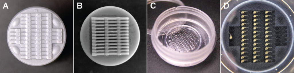
Figure 1 Stamping models and mounted embryos. (A-B) Stamp surfaces of (A) Lateral mounting and (B) Dorsal mounting stamp (C) Ready-to-use agarose imprint with 44 µ-wells. (D) Mounted embryos ready for imaging.
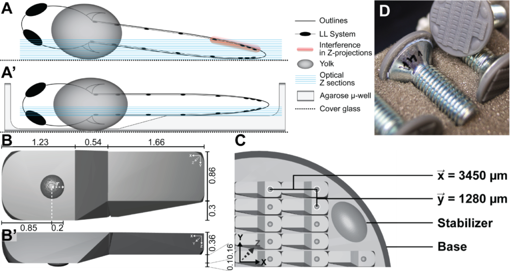
Figure 2 Stamp and µ-well proportions (A, A’) Schemes showing a zebrafish embryo mounted without µ-well (A) and with µ-well (A’). (B, B’) Top-view (B) and orthogonal view (B’) of a single micro well. (C) Design and dimensions of the stamp. The base connects all parts and fits exactly a 20 mm dish. and are the distances in X and Y. Stabilizers were introduced as cornerstones and to make the structure more rigid. (D) For better handling, the stamp is mounted onto a countersunk screw.
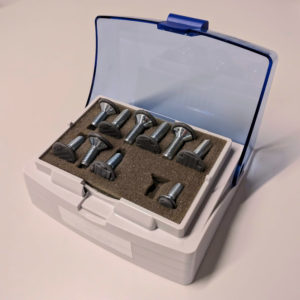
Example Product